
This page has the Life Processes Class 10 chapter — the official NCERT Class 10 Science Chapter 5, as published on ncert.nic.in, running from textbook page 78 to page 99. The official PDF is right below.
Underneath the file, the chapter is explained in plain words: what each section teaches, what the figures show, what the activities prove, and where every exercise is answered.
Download the Life Processes Class 10 chapter PDF
Get the official Life Processes Class 10 NCERT Science Chapter 5 PDF straight from the NCERT website — this is the complete chapter as printed in the textbook, with all its sections, figures, activities and exercises exactly as published, ready to read on any device or save for revision.
The chapter at a glance: what is inside
This section is a map of the chapter: where each topic begins, what the chapter does with it, and what to expect before you start reading.
The chapter opens by asking how we know something is alive (p. 78 of the printed book), then defines life processes as the maintenance processes that keep organisms alive (p. 79). From there it studies the four processes one by one:
- Nutrition — how organisms take in food, pages 80 to 86 (sections 5.2.1 to 5.2.4).
- Respiration — how glucose is broken down for energy, pages 86 to 90.
- Transportation — how materials move around the body, pages 91 to 95.
- Excretion — how wastes are removed, pages 96 to 98.
The book numbers these sections 5.1 to 5.5, and experiment activities appear throughout the chapter — each one demonstrates a specific point about the process being discussed. The chapter closes with the ‘What you have learnt’ summary and the exercise block on pages 98 and 99.
For the exact counts — of sections, figures, activities and questions — the chapter table on this page lists them, and it makes a useful checklist to mark off as you finish each part.
The big idea: what makes something alive
This section is for the chapter’s opening question, because the whole chapter’s logic follows from how it answers ‘what is alive?’. Once you hold this idea, every later process makes sense.
The chapter’s first move is to knock down the easy answer. Visible movement is not enough to define life: a sleeping dog or a cow chewing cud is alive, and so is a plant that is not visibly growing (NCERT, p. 78).
The real evidence of life, the book argues, is molecular movement — invisible changes happening inside cells. This is why viruses, which show no molecular movement until they infect a cell, sit in a grey zone over whether they are alive (NCERT, p. 78).
Why must molecules keep moving? Living bodies are highly ordered structures, and order constantly tends to break down under the effects of the environment. If order breaks, the organism dies. So every living thing must keep repairing and maintaining itself, and that maintenance job is what the chapter calls life processes (NCERT, p. 79).
Maintenance costs energy and raw material, both of which must come from outside the body. That forces a second design problem: in multicellular organisms, not every cell touches the environment. Simple diffusion cannot supply all the cells of a human body.
So food and oxygen are taken up at specialised places in the body, and the body needs a transportation system to deliver them and an excretory system to remove the by-products (NCERT, pp. 79-80). That is the design logic behind the four processes the rest of the chapter studies: nutrition, respiration, transportation and excretion.
Key concepts: the four life processes explained
This section walks through the chapter’s content process by process, in the order the book presents them. Each subsection below matches a numbered section of the textbook, so you can read alongside your book easily.
Nutrition: how organisms take in food
Nutrition is how an organism brings in the energy and raw materials it needs for maintenance and growth. The chapter splits it into two modes. Autotrophs — green plants and some bacteria — build food from simple inorganic materials, carbon dioxide and water, using an external energy source, the Sun (NCERT, p. 80).
Heterotrophs — animals and fungi — depend directly or indirectly on autotrophs for complex food, and use enzymes as bio-catalysts to break it down (NCERT, p. 80).
Photosynthesis is the autotroph’s way of converting carbon dioxide and water into carbohydrates using sunlight and chlorophyll (NCERT, p. 80). The equation the book gives on page 82 is worth reading symbol by symbol:
\[ 6\text{CO}_2 + 12\text{H}_2\text{O} \xrightarrow[\text{Sunlight}]{\text{Chlorophyll}} \text{C}_6\text{H}_{12}\text{O}_6 + 6\text{O}_2 + 6\text{H}_2\text{O} \]
Six molecules of carbon dioxide combine with twelve of water to make one glucose molecule, six molecules of oxygen, and six leftover water molecules. The detail students drop: twelve water molecules go in but only six come out — the other six are split.
Their hydrogen goes into the glucose while the oxygen is released. The reaction needs both chlorophyll and sunlight; that is why leaves are green — chlorophyll is the pigment that absorbs light energy.
Photosynthesis happens in three events (NCERT, pp. 81-82): chlorophyll absorbs light energy; light energy is converted to chemical energy and water is split into hydrogen and oxygen; and carbon dioxide is reduced to carbohydrates.
These steps need not happen one after the other — desert plants take in carbon dioxide at night and use it during the day when sunlight is available (NCERT, p. 82).
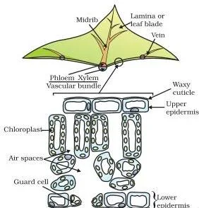
The cross-section in Figure 5.1 shows where photosynthesis happens. Some cells contain green dots — these are the chloroplasts, organelles that hold chlorophyll (NCERT, p. 82).
This is what you actually see under the microscope when you look at that cross-section: green dots scattered in the cells, each one a chloroplast full of chlorophyll. Photosynthesis can only happen where chlorophyll and light meet, which is why these organelles sit in the leaf’s light-catching cells.
The plant takes in carbon dioxide through stomata, tiny pores on the leaf surface. Each pore is bordered by two guard cells. When water flows into the guard cells they swell and the pore opens; when they shrink, the pore closes (Figure 5.3, NCERT, pp. 82-83).
Plants close the pores when they do not need carbon dioxide for photosynthesis, because large amounts of water can also be lost through them.
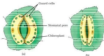
In Figure 5.3, the left pore is open for gas exchange; the right one is closed to save water. That pairing — take in carbon dioxide when needed, close up when not — is the plant’s water-saving compromise.
Beyond carbon dioxide and water, plants need soil minerals. Roots take up water and minerals like nitrogen, phosphorus, iron and magnesium. Nitrogen is essential for making proteins; it is absorbed as inorganic nitrates or nitrites, or as organic compounds that bacteria prepare from atmospheric nitrogen (NCERT, p. 83).
Heterotrophic nutrition comes in more than one strategy (NCERT, p. 83). Some organisms, like the fungi bread mould, yeast and mushrooms, break food down outside the body and then absorb the simpler products. Others take whole food in and break it down inside.
Parasites — cuscuta, ticks, lice, leeches, tapeworms — take nutrition from a plant or animal without killing it. In single-celled organisms the method depends on body design. Amoeba uses temporary finger-like extensions of the cell surface that fuse over the food particle to form a food vacuole; digestion happens inside the vacuole and the undigested material is thrown out (NCERT, pp. 83-84).
Paramoecium, which has a definite shape, moves food to a fixed spot using the cilia that cover its surface (NCERT, p. 84).
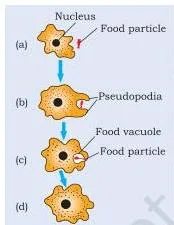
The Amoeba figure shows the food vacuole forming: the cell reaches out, surrounds the particle, and seals it inside. Everything that follows — digestion, diffusion into the cytoplasm, ejection of the leftovers — happens around that vacuole.
Human digestion runs along the alimentary canal, a long tube from mouth to anus with specialised regions (Figure 5.6, NCERT, p. 84). Follow the food part by part:
- Mouth. Teeth crush food; saliva from the salivary glands contains salivary amylase, which breaks starch down into simple sugar (NCERT, p. 84).
- Oesophagus. Rhythmic muscle contractions called peristaltic movements push the food forward (NCERT, p. 84).
- Stomach. Gastric glands release hydrochloric acid, the protein-digesting enzyme pepsin, and mucus. The acid makes the medium acidic so pepsin can act; its other function, the answer to the book’s question, is killing bacteria in food. Mucus protects the stomach lining (NCERT, p. 85).
- Small intestine. The longest part of the canal, coiled to fit a compact space. Bile from the liver makes the food alkaline for the pancreatic enzymes, and bile salts break large fat globules into smaller ones — the book compares this to the emulsifying action of soap on dirt (NCERT, p. 85).
- Completing digestion. Pancreatic juice supplies trypsin for proteins and lipase for fats; intestinal juice finishes the job, converting proteins to amino acids, carbohydrates to glucose, and fats to fatty acids and glycerol (NCERT, p. 85).
- Large intestine. Its wall absorbs water from the unabsorbed material; the anal sphincter regulates removal from the body (NCERT, p. 85).
Memory aid: the organs in order spell M-O-S-S-L-A — Mouth, Oesophagus, Stomach, Small intestine, Large intestine, Anus. Food always moves in that order, with the small intestine doing most of the digesting and the large intestine mopping up water.
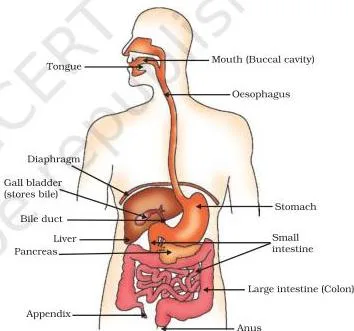
Trace the tube in Figure 5.6 from top to bottom and you are tracing the path of food. Each region is specialised, and the sphincter at the stomach’s exit releases food in small amounts to give the small intestine time to finish the job (NCERT, p. 85).
Absorption happens in the small intestine, whose inner lining has finger-like projections called villi. The villi increase the surface area for absorption and are richly supplied with blood vessels, which carry the absorbed food to every cell (NCERT, p. 85).
Herbivores that eat grass need a longer small intestine because cellulose is harder to digest; carnivores like tigers digest meat more easily and have a shorter one (NCERT, p. 85).
One health note the chapter adds: dental caries, or tooth decay. Bacteria acting on sugars produce acids that soften and demineralise the enamel. The bacterial mass plus food particles forms plaque, which blocks saliva from neutralising the acid — which is why brushing after eating matters (NCERT, p. 85).
Checkpoint: the five in-text questions on page 86 ask you to compare autotrophic and heterotrophic nutrition, list where plants get each raw material, state the role of stomach acid, and explain how the small intestine absorbs food. If you can answer those, this section is done.
Respiration: getting energy from glucose
Respiration is the process by which the energy in food is released inside cells. It is not the same as breathing — the chapter defines respiration as acquiring oxygen and using it in the breakdown of food for cellular needs (NCERT, p. 79). The breakdown itself happens in steps.
In all organisms the first step is the same: glucose, a six-carbon molecule, is broken in the cytoplasm into pyruvate, a three-carbon molecule (NCERT, pp. 86-87). From pyruvate, the next step splits into three pathways, shown in Figure 5.8:
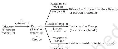
- Yeast (anaerobic). Pyruvate becomes ethanol and carbon dioxide without oxygen — fermentation (NCERT, p. 87).
- Mitochondria (aerobic). Pyruvate is broken down using oxygen into carbon dioxide and water. The energy release here is far greater than in the anaerobic process (NCERT, p. 87).
- Muscle cells short of oxygen. Pyruvate becomes lactic acid, a three-carbon molecule; its build-up during sudden activity causes cramps (NCERT, p. 87).
Read Figure 5.8 as a flow chart: one start (glucose in the cytoplasm), one junction (pyruvate), and three exits. The detail that matters in answers is the location — cytoplasm for the first step, mitochondria for the aerobic one — and the energy difference.
The energy released in respiration is not used directly. The cell uses it to build ATP (adenosine triphosphate) from ADP and inorganic phosphate (NCERT, p. 88):
\[ \text{ADP} + \text{P} \xrightarrow{\text{Energy}} \text{ATP} \]
When the terminal phosphate link in ATP is broken using water, about 30.5 kJ per mole of energy is released to drive the cell’s reactions (NCERT, pp. 87-88). The chapter’s comparison is a battery: one stored form of energy that can power muscle contraction, protein synthesis, nervous impulses and many other activities.
ATP is made on demand, not stored as a long-term fuel.
Plants exchange gases through stomata and large inter-cellular spaces, by diffusion. By day, the carbon dioxide made in respiration is used up in photosynthesis, so no carbon dioxide is released and oxygen is the main gas given out.
At night, when photosynthesis stops, carbon dioxide is the gas being eliminated (NCERT, p. 88). This is the point students confuse — plants respire all the time, they only photosynthesise in light.
Aquatic organisms face a problem terrestrial ones do not: dissolved oxygen is far less plentiful than atmospheric oxygen. So fish breathe fast — they take water in through the mouth and force it past the gills, where dissolved oxygen is picked up by blood (Activity 5.6, NCERT, p. 89).
Human breathing takes air in through the nostrils, where fine hairs and mucus filter it. Rings of cartilage keep the throat passage from collapsing. Inside the lungs the passage divides into smaller tubes ending in balloon-like alveoli, whose walls carry an extensive network of blood vessels (NCERT, pp. 89-90).
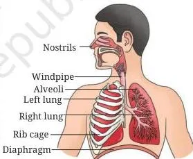
Trace the path in Figure 5.9: nostrils, throat, lungs, alveoli. The alveoli are where exchange happens, and the residual volume of air kept in the lungs between breaths means gas exchange continues even during the pause in breathing (NCERT, p. 90).
Why so much surface? If the alveolar surface were spread flat it would cover about 80 square metres (NCERT, p. 90). The same logic explains haemoglobin: diffusion alone would take an estimated three years for oxygen to reach the toes.
The respiratory pigment haemoglobin in red blood cells has a very high affinity for oxygen and carries it to tissue that needs it; carbon dioxide, being more soluble in water, is mostly carried dissolved in the blood (NCERT, p. 90).
The book adds a warning with a biological reason. Smoking destroys the hair-like cilia in the upper respiratory tract that remove germs and dust from inhaled air. Once they are gone, that material reaches the lungs and can cause infection, cough and even lung cancer (NCERT, p. 90).
Checkpoint: the four in-text questions on page 90 ask about the terrestrial advantage in obtaining oxygen, the ways glucose is oxidised, how oxygen and carbon dioxide are transported, and how the lungs maximise exchange area.
Transportation: moving materials around the body
Transportation exists because food and oxygen enter at one place but every cell needs them. The human transport system has three parts: a pump (the heart), tubes (blood vessels), and a repair system (platelets).
Blood itself is the medium. Its fluid part, plasma, carries food, carbon dioxide and nitrogenous wastes in dissolved form; red blood cells carry oxygen using haemoglobin (NCERT, p. 91).
The heart is a muscular organ about the size of a fist, with four chambers. Follow the blood’s path (NCERT, pp. 91-92, Figure 5.10):
- Oxygen-rich blood from the lungs enters the left atrium, passes to the left ventricle, which pumps it to the rest of the body.
- Deoxygenated blood from the body enters the right atrium, passes to the right ventricle, which pumps it to the lungs for oxygenation.
- Valves between chambers stop blood flowing backwards, and the ventricles have thicker muscular walls than the atria because they pump blood to distant organs.
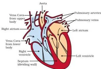
In Figure 5.10, the side matters: the left chambers handle oxygenated blood, the right chambers handle deoxygenated blood. This separation stops the two streams mixing.
Memory aid: the right side routes blood to the lungs, the left side routes it to the rest of the body. When you draw the heart, label the two sides accordingly.
Because the blood passes through the heart twice in one complete cycle, this is called double circulation (NCERT, p. 92, Figure 5.11). Separating oxygenated and deoxygenated blood gives a highly efficient oxygen supply — essential for birds and mammals, which constantly use energy to keep their body temperature steady.
Amphibians and many reptiles have three-chambered hearts and tolerate some mixing; fish have two-chambered hearts and pass blood through the heart only once per cycle (NCERT, p. 92).
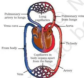
Figure 5.11 shows the two loops of double circulation: the loop to the lungs, where deoxygenated blood drops carbon dioxide and picks up oxygen, and the loop around the body, where oxygenated blood delivers oxygen and collects carbon dioxide. The blood goes through the heart twice — hence ‘double’.
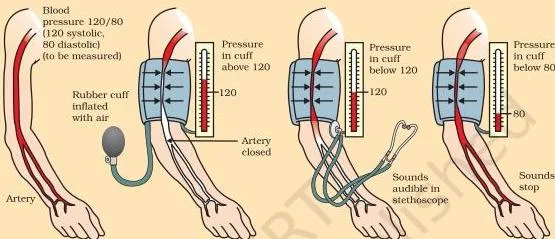
Blood pressure is the force blood exerts against the walls of the vessels (NCERT, p. 92). It is higher in arteries than in veins. The pressure during ventricular contraction, systolic, is normally about 120 mm of Hg; during relaxation, diastolic, about 80 mm of Hg.
Blood pressure is measured with a sphygmomanometer. High blood pressure, or hypertension, comes from constricted arterioles and can rupture an artery, causing internal bleeding (NCERT, pp. 92-93).
The vessel types follow from their jobs (NCERT, p. 93). Arteries carry blood away from the heart under high pressure, so their walls are thick and elastic. Veins return blood to the heart, so they have valves that keep blood flowing one way.
Capillaries are one cell thick, and it is across their thin walls that materials exchange between blood and cells. Platelets plug leaks in this network by helping blood clot at injury points.
A second fluid also transports. Lymph, or tissue fluid, forms when plasma, proteins and blood cells escape through capillary pores into the intercellular spaces. It is colourless and contains less protein than plasma, and it carries absorbed fat from the intestine and drains excess fluid from the tissues back into the blood (NCERT, p. 93).
Plants need a transport system too, but a slower one. Because plants do not move and many of their tissues are dead cells, they have low energy needs and can use slow transport over long distances (NCERT, p. 93).
The vascular tissue has two independent pathways. Xylem carries water and minerals from the roots; phloem carries the products of photosynthesis from the leaves to the rest of the plant (NCERT, p. 94).
Transpiration pull is the major driving force for water movement by day. Evaporation of water from leaf cells creates a suction that pulls water up from the roots; the loss of water vapour from aerial parts is transpiration, and it also helps regulate temperature. At night, when stomata are closed, root pressure becomes more important (NCERT, p. 95, Figure 5.12).
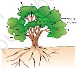
Figure 5.12 shows the whole route: water enters at the roots, climbs the xylem, and is drawn upward by the suction from the leaves. That is why the pull is called transpiration pull — the leaves do the pulling.
Food transport, or translocation, works in the phloem: sucrose and amino acids move in sieve tubes with the help of adjacent companion cells, in both upward and downward directions. Unlike xylem transport, this uses energy from ATP — phloem moves material according to the plant’s needs, such as sugar stored in roots moving to buds in spring (NCERT, pp. 94-95).
| Feature | Xylem | Phloem |
|---|---|---|
| What it carries | Water and minerals from the soil | Products of photosynthesis — sucrose, amino acids and other substances |
| Direction | Upward, from roots to leaves | Both upward and downward, to storage and growing organs |
| Energy use | Largely explained by simple physical forces (root pressure, transpiration pull) | Uses energy from ATP; an active process |
| Tissue involved | Vessels and tracheids forming continuous water-conducting channels | Sieve tubes with adjacent companion cells |
| NCERT page | pp. 93-95 | pp. 94-95 |
Checkpoint: the five in-text questions on page 95 ask for the components of the human and plant transport systems, why mammals and birds separate oxygenated and deoxygenated blood, and how water, minerals and food move in plants.
Excretion: removing the waste
Excretion is the removal of harmful metabolic wastes. Chemical reactions that keep us alive also produce by-products that are useless or even harmful — especially nitrogenous wastes like urea and uric acid. Many unicellular organisms simply diffuse these out through the body surface; complex organisms use specialised organs (NCERT, p. 96).
The human excretory system has a pair of kidneys in the abdomen, one on each side of the backbone, a pair of ureters, a urinary bladder and a urethra (NCERT, p. 96, Figure 5.13).
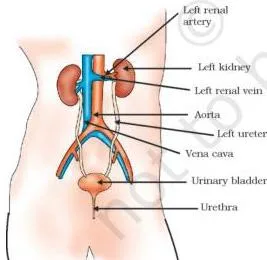
Figure 5.13 gives the layout and the flow: urine is made in the kidneys, travels down the ureters, is stored in the bladder, and leaves through the urethra. The bladder is muscular and under nervous control, which is why we can usually hold the urge to urinate (NCERT, p. 97).
The kidney’s job is to filter nitrogenous waste out of the blood. The basic filtration unit is the nephron — a cluster of thin-walled capillaries associated with the cup-shaped end of a coiled tube, Bowman’s capsule (NCERT, pp. 96-97, Figure 5.14).
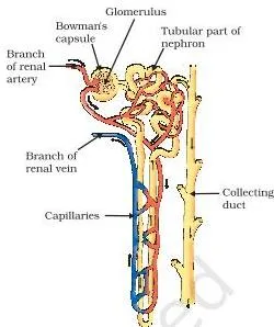
In Figure 5.14, two jobs are clearly separated. At the top, filtration: the capillary cluster pushes an initial filtrate into Bowman’s capsule. Along the coiled tube, selective reabsorption takes back glucose, amino acids, salts and most of the water, depending on how much excess water the body has and how much dissolved waste must be excreted.
What remains flows down the tube as urine (NCERT, pp. 96-97).
Worked example — how much filtrate the body reabsorbs.
The chapter gives the numbers on page 97: a healthy adult’s kidneys make about 180 litres of initial filtrate daily, but only a litre or two are passed out as urine.
Step 1: Take urine output as 1.5 litres.
Reabsorbed filtrate = 180 − 1.5 = 178.5 litres.
\[ \frac{178.5}{180} \times 100 = 99.17\% \approx 99\% \]
Final answer: about 99 percent of the daily filtrate is reabsorbed before urine is formed — which is why the kidneys can maintain blood composition without draining the body.
When kidneys fail, wastes accumulate and can be fatal. An artificial kidney (hemodialysis) removes nitrogenous wastes by dialysis: blood passes through tubes with semi-permeable linings, suspended in a tank of dialysing fluid that has the same osmotic pressure as blood but no nitrogenous wastes.
The wastes diffuse out and the purified blood returns to the body. The key difference from a real kidney: dialysis involves no reabsorption (NCERT, p. 97).
A short note closes the human part: organ donation — kidneys, part of a liver, corneas, heart and other organs — can save lives, and anyone, regardless of age or gender, can be a donor (NCERT, pp. 97-98).
Plants use completely different strategies (NCERT, p. 98). Oxygen itself is a by-product of photosynthesis. Excess water leaves through transpiration. Other wastes are stored in cellular vacuoles, or in leaves that then fall off, or as resins and gums in old xylem. And some waste substances are excreted into the soil around the plant.
Checkpoint: the three in-text questions on page 97 ask you to describe the structure and functioning of nephrons, list how plants get rid of excretory products, and explain how urine amount is regulated.
The activities in the chapter and what each one proves
This section collects the chapter’s experiment activities — useful for practical work and viva preparation. Each row says what you do and the conclusion the experiment supports.
Two experiments in the nutrition section are worth seeing as a pair, because they isolate one factor at a time. Activity 5.1 tests whether chlorophyll is needed for photosynthesis.
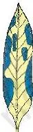
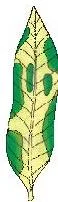
In Figure 5.2, the plant is kept in the dark for three days to use up its starch, then returned to sunlight. A leaf is decolourised with boiling alcohol and tested with iodine. The green areas — which had chlorophyll — turn blue-black, showing starch was made there. The non-green areas stay unchanged.
Conclusion: chlorophyll is essential for photosynthesis (NCERT, p. 82). Activity 5.2 does the same trick for carbon dioxide.
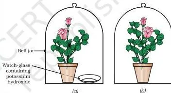
In Figure 5.4, both set-ups are sealed with vaseline. Set-up (a) holds potassium hydroxide, which absorbs carbon dioxide; set-up (b) is the control. After sunlight, the leaf from the jar without potassium hydroxide shows starch; the leaf from the jar with it shows little or none.
Conclusion: carbon dioxide is needed for photosynthesis (NCERT, p. 83). Here is the full list of the chapter’s activities:
| Activity (page) | What you do | What it proves |
|---|---|---|
| 5.1 (p. 82) | Keep a variegated plant in the dark to use up starch, then in sunlight; test a leaf with iodine after decolourising in alcohol. | Chlorophyll is essential for photosynthesis — only the green areas turn blue-black. |
| 5.2 (p. 83) | Two plants in sealed bell jars, one with potassium hydroxide to absorb carbon dioxide; test leaves for starch after sunlight. | Carbon dioxide is essential for photosynthesis — the leaf from the jar with potassium hydroxide shows little or no starch. |
| 5.3 (p. 85) | Add saliva to a starch solution, wait 20-30 minutes, then test with iodine. | Salivary amylase in saliva breaks starch down to sugar — that tube shows no blue-black colour. |
| 5.4 (p. 87) | Pass ordinary air and exhaled air through lime water and compare how fast they turn milky. | Exhaled air contains more carbon dioxide — it turns lime water milky faster. |
| 5.5 (p. 87) | Add yeast to a sugar solution; bubble the gas produced through lime water. | Fermentation releases carbon dioxide — the lime water turns milky. |
| 5.6 (p. 89) | Watch fish open and close their mouths and gill-slits; count how often, and compare with your own breathing. | Aquatic animals breathe faster because the oxygen dissolved in water is comparatively low. |
| 5.7 (p. 91) | Visit a health centre and find the normal haemoglobin range; compare children and adults, men and women. | Normal haemoglobin differs with age and between men and women. |
| 5.8 (p. 95) | Cover a potted plant and a bare stick separately in bright sunlight, pot soil sealed with plastic. | Transpiration releases water vapour — droplets appear inside the cover over the plant, not the stick. |
How to read the chapter’s key diagrams
This section treats the chapter’s most information-rich diagrams as drawings you must be able to read and reproduce — the glucose pathway, the heart with double circulation, and the nephron. The same figures appear in the concept sections; here the focus is on what to draw and what to label.
The break-down of glucose (Figure 5.8)
Read it as a flow chart. Start with glucose, six carbons, labelled ‘in the cytoplasm’. Below it, pyruvate, three carbons — also in the cytoplasm. From pyruvate, three arrows:
- to ethanol + carbon dioxide — yeast, anaerobic (fermentation);
- to carbon dioxide + water — in the mitochondria, aerobic, releasing far more energy;
- to lactic acid — in muscle cells when oxygen is short, the cause of cramps.
Reproduction checklist: draw boxes for glucose and pyruvate, connect them, then draw the three branches and write the location next to each — cytoplasm, mitochondria, cytoplasm. Labelling the locations is what makes the answer complete.
The heart and double circulation (Figures 5.10 and 5.11)
Read Figure 5.10 before drawing it. Fix in your mind the four chambers and which blood each one holds:
- Left atrium — receives oxygen-rich blood from the lungs.
- Left ventricle — pumps that blood to the body.
- Right atrium — receives deoxygenated blood from the body.
- Right ventricle — pumps it to the lungs.
The order to trace: lungs → left atrium → left ventricle → body, and body → right atrium → right ventricle → lungs. The double circulation in Figure 5.11 is exactly those two loops, with the heart at the centre of both — one loop to the lungs, one loop around the body.
Reproduction checklist for the heart: draw four chambers, label left atrium and ventricle on one side and right atrium and ventricle on the other; make the ventricular wall thicker than the atrial wall; add valves between atria and ventricles; label the left side ‘oxygenated’ and the right side ‘deoxygenated’.
The nephron (Figure 5.14)
Read the nephron as two jobs in one diagram. At the top: the cluster of thin-walled capillaries inside Bowman’s capsule — this is where filtration happens, producing the initial filtrate. Below it: the coiled tube — this is where selective reabsorption of glucose, amino acids, salts and water happens. The flow runs one way: filtrate in, urine out.
Reproduction checklist: draw the capillary cluster, cup-shaped Bowman’s capsule around it, the coiled tubule, and an arrow showing urine leaving; label ‘filtration’ at the capsule and ‘reabsorption’ along the tube; note that nephrons are packed close together in each kidney.
Definitions you will need in your answers
This glossary holds the chapter’s key terms, defined the way you need them in an answer. Each entry carries the textbook page where the term is used.
| Term | Plain definition | NCERT page |
|---|---|---|
| Life processes | The maintenance functions that keep an organism alive — nutrition, respiration, transportation and excretion. | p. 79 |
| Nutrition | The process of transferring a source of energy (food) from outside the body to the inside. | p. 79 |
| Autotrophic nutrition | Building complex food from simple inorganic materials — carbon dioxide and water — using sunlight and chlorophyll. | pp. 80-81 |
| Heterotrophic nutrition | Taking in complex food prepared by other organisms and breaking it down with enzymes. | p. 80 |
| Photosynthesis | The process by which autotrophs convert carbon dioxide and water into carbohydrates using sunlight and chlorophyll. | pp. 80-81 |
| Enzyme | A bio-catalyst that breaks down complex food substances into simpler ones. | p. 80 |
| Peristalsis | The rhythmic contractions of the gut lining that push food along the alimentary canal. | p. 84 |
| Villi | Finger-like projections of the small intestine lining that increase the surface area for absorbing digested food. | p. 85 |
| Aerobic respiration | Breakdown of pyruvate using oxygen, in the mitochondria, releasing a great deal of energy. | p. 87 |
| Anaerobic respiration | Breakdown of pyruvate without oxygen, as in yeast fermentation, releasing less energy. | p. 87 |
| ATP | Adenosine triphosphate — the energy currency of the cell, made from ADP and phosphate using energy from respiration. | pp. 87-88 |
| Blood pressure | The force blood exerts against the wall of a vessel; systolic about 120 mm of Hg and diastolic about 80 mm of Hg in a normal adult. | pp. 92-93 |
| Double circulation | The passage of blood through the heart twice during one complete cycle of the body. | p. 92 |
| Transpiration | Loss of water as vapour from the aerial parts of a plant; it pulls water up the xylem and regulates temperature. | p. 95 |
| Translocation | Transport of soluble products of photosynthesis in the phloem, in upward and downward directions. | p. 95 |
| Excretion | Removal of harmful metabolic wastes, especially nitrogenous wastes like urea and uric acid. | p. 96 |
| Dialysis | Removal of nitrogenous wastes from blood in an artificial kidney by diffusion across a semi-permeable lining, without reabsorption. | p. 97 |
Mistakes students make in this chapter, corrected
This chapter has a handful of confusions that show up in students’ answers again and again. Each row names the mistake, gives the correct rule, and tells you how to check your answer in the book.
| Mistake | Correct rule | How to check your answer |
|---|---|---|
| ‘Plants photosynthesise at night and respire only by day.’ | Plants respire all the time. By day the carbon dioxide they produce is used up in photosynthesis, so none is released; at night carbon dioxide is eliminated. | p. 88 — the gas-exchange paragraph explains day and night separately. |
| ‘The left side of the heart carries deoxygenated blood.’ | The left chambers handle oxygenated blood coming back from the lungs; the right chambers handle deoxygenated blood. | pp. 91-92 — trace the blood path in Figure 5.10. |
| ‘Breathing and respiration are the same thing.’ | Breathing is the mechanical taking in and letting out of air; respiration is acquiring oxygen and using it to break down food for cellular needs. | p. 79 — the definition of respiration. |
| ‘The stomach digests fats.’ | Fats are digested in the small intestine, where bile salts emulsify them and lipase breaks them into fatty acids and glycerol. | p. 85 — the small intestine paragraph. |
| ‘The only job of hydrochloric acid in the stomach is to make the medium acidic.’ | It makes the medium acidic for pepsin — and its other function, the book’s own question, is killing bacteria in food. | p. 85 — the gastric glands paragraph. |
| ‘Xylem and phloem both carry food.’ | Xylem carries water and minerals upward; phloem carries food in both directions using energy from ATP. | pp. 93-95 — the xylem/phloem comparison table above. |
| ‘The photosynthesis equation has six water molecules on both sides.’ | Twelve water molecules go in, six come out; the other six are split, their hydrogen used to build glucose. | p. 82 — balance the equation yourself. |
| ‘Anaerobic respiration releases more energy.’ | Aerobic respiration releases far more energy than anaerobic respiration. | p. 87 — the respiration pathway text. |
| ‘Urine is just filtered blood.’ | Filtration is followed by selective reabsorption; about 180 litres are filtered daily but only one or two litres leave as urine. | p. 97 — the dialysis note gives the 180-litre figure. |
| ‘Every artery carries oxygenated blood.’ | The vessel from the right ventricle to the lungs carries deoxygenated blood — the exception to the rule. | p. 92 — the heart path description. |
Where every chapter exercise is answered
When you finish the chapter, test yourself with its own questions. This map tells you where each answer lives in the chapter.
| Exercise | Question | Where the answer is | What a complete answer covers |
|---|---|---|---|
| 1 | Kidneys are part of which system? | Section 5.5 (p. 96) | Excretion. |
| 2 | What is the xylem responsible for? | Section 5.4.2 (p. 94) | Transport of water. |
| 3 | What does autotrophic nutrition require? | Section 5.2.1 (p. 81) | Carbon dioxide and water, chlorophyll, and sunlight — all of the above. |
| 4 | Where is pyruvate broken down with oxygen? | Respiration section (p. 87) | In the mitochondria. |
| 5 | How are fats digested and where? | Section 5.2.4 (p. 85) | Bile salts emulsify fat globules; lipase breaks fats to fatty acids and glycerol in the small intestine. |
| 6 | Role of saliva in digestion | Section 5.2.4 (p. 84) | Salivary amylase breaks starch into simple sugar. |
| 7 | Conditions and by-products of autotrophic nutrition | Section 5.2.1 (pp. 81-82) | Conditions: carbon dioxide, water, sunlight, chlorophyll; by-products: oxygen (and water). |
| 8 | Aerobic vs anaerobic respiration; organisms using anaerobic mode | Figure 5.8 (p. 87) | Oxygen use, location, energy yield; yeast as the example organism for fermentation. |
| 9 | How alveoli maximise gas exchange | Respiration section (p. 90) | Balloon-like alveoli, extensive network of blood vessels, residual air keeps exchange continuous, large surface area (about 80 m²). |
| 10 | Consequences of haemoglobin deficiency | Respiration section (p. 90) | The body cannot carry enough oxygen to the tissues, because haemoglobin is the oxygen-carrying pigment of red blood cells. |
| 11 | Describe double circulation; why is it necessary | Transportation section (p. 92) | Blood passes through the heart twice per cycle; separation of oxygenated and deoxygenated blood gives a highly efficient oxygen supply for animals that maintain body temperature. |
| 12 | Differences between xylem and phloem transport | Plant transport (pp. 94-95) | What each carries, direction, energy use — use the xylem/phloem table above. |
| 13 | Compare alveoli and nephrons | Respiration (p. 90) and excretion (pp. 96-97) | Structure-function: both are thin-walled surfaces with a rich blood supply; alveoli exchange gases, nephrons filter blood and selectively reabsorb. |
Also use the in-text question sets after each section — on pages 80, 86, 90, 95 and 97 — as checkpoints as you read, and the chapter’s ‘What you have learnt’ on pages 98-99 as a condensed answer key for most of the above.
One honest note: textbook contents and the examinable syllabus are not always identical — check the current official syllabus for what is examinable this session.
The chapter’s takeaways in one screen
This is the chapter’s own summary, ‘What you have learnt’ (pp. 98-99), restated in plainer words for last-minute revision.
- Movement — including invisible molecular movement — is the indication of life.
- Maintaining life needs four things: nutrition, respiration, transport of materials, and excretion of wastes.
- Autotrophic nutrition makes complex food from simple inorganic materials using sunlight; heterotrophic nutrition uses food prepared by other organisms.
- In human beings, food is broken down step by step along the alimentary canal, and the digested food is absorbed in the small intestine and sent to all cells.
- Respiration breaks down organic compounds such as glucose to provide energy in the form of ATP, which then drives other cellular reactions.
- Aerobic respiration makes more energy available to the organism than anaerobic respiration.
- In human beings, the circulatory system — heart, blood and blood vessels — transports oxygen, carbon dioxide, food and excretory products.
- In plants, the vascular tissue — xylem and phloem — transports water, minerals and food.
- Nephrons in the kidneys remove soluble nitrogenous waste from the blood.
- Plants store wastes in vacuoles, as gums and resins, in leaves that fall off, or excrete them into the surrounding soil.
Where Life Processes sits in the Class 10 Science book
Life Processes is Chapter 5 of the NCERT Class 10 Science book. The natural next read is Chapter 6, Control and Coordination, which explains how the body controls and coordinates these very processes.
The chemistry behind the digestion section sits in Chapter 4, Carbon and its Compounds — the book itself points back to it when it compares bile salts to the emulsifying action of soap (NCERT, p. 85).
For the wider syllabus, see the Class 10 notes hub or start from the CBSE notes index.
Sources and data verification
This listing is maintained for the 2026-27 academic session using the NCERT textbook information available to us. NCERT remains the authority for confirming the latest edition.
The page references on this page come from the current NCERT Class 10 Science textbook, Chapter 5 (Life Processes), rationalised edition, as published on ncert.nic.in. Figure pages follow the printed book, with figures cited on textbook pages 82 to 97.
NCERT settles the textbook, its editions and its PDFs; CBSE settles the examinable syllabus. This page is maintained for the current academic session using the NCERT information available to us, and it covers this single chapter — not the whole Class 10 Science book.
| What the chapter holds | Count | Where it is used |
|---|---|---|
| Printed pages | 21 | |
| Sections in the chapter | 12 | |
| Figures with NCERT captions | 15 | |
| Exercise questions | 13 | answered in our NCERT Solutions |
| In-text questions | 21 | |
| Activities | 8 | |
| Official NCERT PDF | Download the chapter PDF | the chapter exactly as NCERT publishes it |
Reference: NCERT Class 10 Science textbook, chapter 5, official edition on ncert.nic.in.
Common questions about Life Processes, answered
Why is diffusion not enough to meet the oxygen requirements of a human being?
Because in a multicellular body not all cells touch the environment. Oxygen is taken in at one place — the lungs — and has to be transported to every cell, so simple diffusion cannot supply all the cells. That is why the body needs a transport system (NCERT, p. 80).
What is the difference between breathing and respiration?
Breathing is the mechanical taking in of air and letting it out. Respiration, as the chapter defines it, is acquiring oxygen and using it in the breakdown of food for cellular needs — a chemical process inside cells (NCERT, pp. 79, 87).
Why is it necessary to separate oxygenated and deoxygenated blood in mammals and birds?
Separation gives a highly efficient supply of oxygen to the body, which matters for animals that constantly use energy to maintain a steady body temperature. This is also the chapter’s own exercise 11 (NCERT, p. 92).
How much blood filtrate do the kidneys make each day and how much becomes urine?
A healthy adult’s kidneys filter about 180 litres of initial filtrate daily, but only one or two litres are passed out as urine — the rest is reabsorbed in the kidney tubules. That is roughly 99 percent of the filtrate (NCERT, p. 97).
What is the role of hydrochloric acid in the stomach?
It makes the medium acidic so that the enzyme pepsin can act on proteins. The book asks what other function the acid serves; the answer it is looking for is that the acid kills bacteria that come in with food (NCERT, p. 85).
How do plants get rid of their waste products?
Excess water leaves through transpiration. Other wastes are stored in cellular vacuoles, in leaves that fall off, or as resins and gums in old xylem; some wastes are excreted into the soil around the plant (NCERT, p. 98).
Explore Class 10 Science Books
Related chapters:
- Chemical Reactions and Equations
- Acids, Bases and Salts
- Metals and Non-metals